To date, the atomic structure of more than 80,000 proteins have been solved by X-ray diffraction methods. A key behind the great success of diffractive methods in structural biology is the possibility to grow crystals, consisting of millions of identical proteins. However, such crystals are not always possible to form, especially in insoluble and fragile molecules such as membrane proteins and protein complexes. To overcome this limitation of protein crystallography is of outmost biological importance, and will completely change the field of structural biology. We have predicted, via molecular dynamics simulations, that very intense and short X-ray pulses allows imaging of single molecules without a crystalline periodicity [1] and we have recently succeeded to image a single virus using micron focal and dense X-ray pulses [2]. Using a combination of developments in instrumentation, theory, and data analysis methods we are constantly pushing the limits in non-crystalline diffraction. The final goal of this project is to image 3D atomic structures of single molecules without crystallization.
Our challenges
Submicron aiming
The 100 nm focus achieved at LCLS is necessary in order to get the photon densities needed for single molecule diffraction. Our challenge is to inject single protein molecules (10-100 nm) individually within the submicron focal spot of the X-ray pulses (See sample delivery).
Hit finding
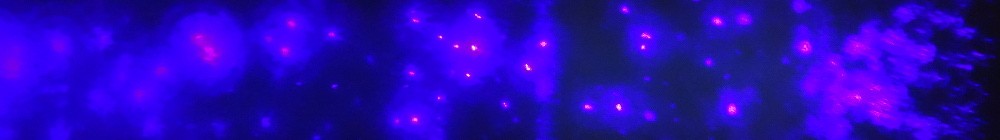
Diffraction from single molecules, even at the most powerful X-ray sources available today, are expected to be very weak. In some cases, the number of diffracted photons will be fewer than those detected from the X-ray beam itself. Still, structural information can be obtained if we can correctly identify when the X-ray beam hits a single molecule and which of the detected photons are likely to arise from the diffraction.
To this end, we are developing “hit finding” methods, where hits are identified via a combination of statistical methods on the diffraction signal and auxiliary ion Time of Flight (iTOF) detection from the sample fragmentation that results from the interaction between sample and X-ray pulse.
3D reconstruction
Individual diffraction patterns give 2D projections of objects. Complete 3D reconstruction of the molecular structure requires alignment and phasing of many such patterns (See reconstruction and phasing algorithms).
Imaging of a single RNA polymerase II
We have performed single molecule imaging on the 15 nm protein complex RNA polymerase II. The eukaryotic RNA polymerase II has fundamental functions in synthesizing mRNA, hnRNA (heterogeneous nuclear RNA) and snRNA (small nuclear RNA). By employing our newly developed hit-finding methods in conjunction with extreme X-ray focusing at the coherent X-ray imaging (CXI) beam line, LCLS, we have managed to identify diffraction from single RNA polymerase II. We now possess the experimental instrumentation and infrastructure required to inject and hit single molecules with a free-electron X-ray pulse as well as identifying the resulting diffraction.
Publications:
1. Neutze, et al., Nature 406: 752-757 (2000). Read Online
2. Seibert, Ekeberg, Maia, et al., Nature 470: 78-81 (2011). Read Online